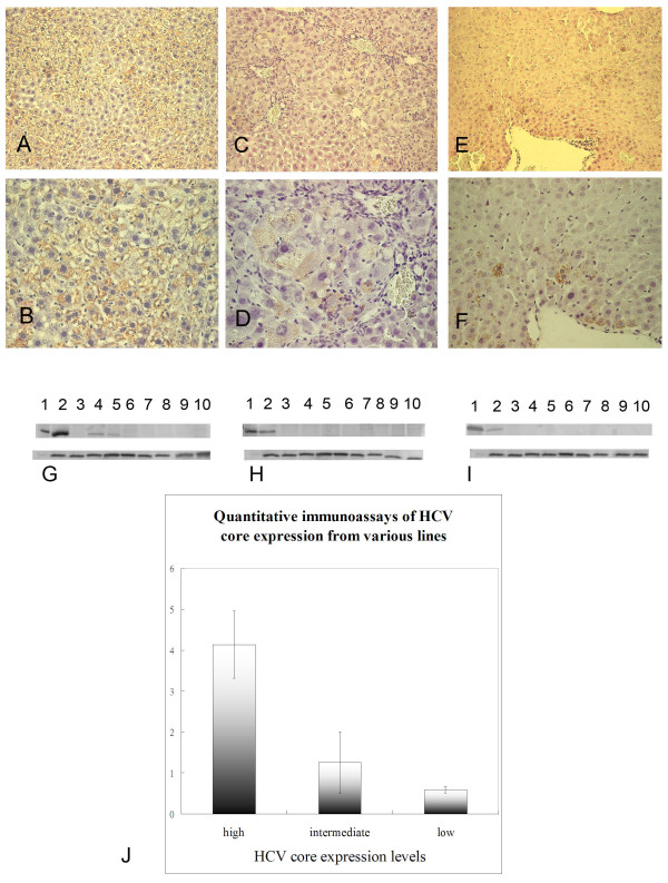
Figure 1.
Immunohistochemical stain for HCV core in the 2-month-old double transgenic mouse (DTM) liver with high (A, B), intermediate (C, D) and low (E, F) HCV core protein expression. The upper region of panels G, H and I depict results of Western blots for HCV core protein extracted from the same livers after two months on control chow (permissive diet). Detection in DTM with high (G), intermediate (H), and low HCV core protein expression (I). Lane 1, HeLa cells transfected with HCV core plasmid (positive control, He); lane 2, liver (li); lane 3, heart (h); lane 4, kidney (k); lane 5, thymus (t); lane 6, omental fat (o); lane 7, lung (lu); lane 8, muscle (m); lane 9, intestine (i); lane 10, skin (s). As a loading control, the same samples were probed for GAPDH (lower panel G, H, and I). (J) Quantitative immunoassays for hepatic HCV core protein from high, intermediate, and low protein-expressing mouse lines. Y-axis, core bands densities (units) acquired from Fluor-S multiimager.

