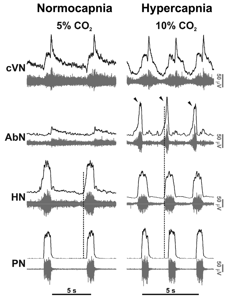
Figure 2. Eupneic pattern of respiratory motor activity in situ.
In control conditions (normocapnia) phrenic (PN) and hypoglossal (HN) have an inspiratory ramp shaped activity envelope whereas abdominal nerve (AbN; lumbar segment 1) exhibits small amplitude post-inspiratory activity. Using 10% carbon dioxide (i.e. 5% above normocapnic conditions), central respiratory drive was raised. This resulted in generation of augmenting expiratory activity in the AbN outflow (Late-E; arrowed) and advanced the onset of pre-inspiratory HN activity relative to PN; the latter indicating reduced airway resistance during both the forced expiration and inspiration. The pattern of PN now showed an abrupt onset in discharge (see Abdala et al. 2009).

