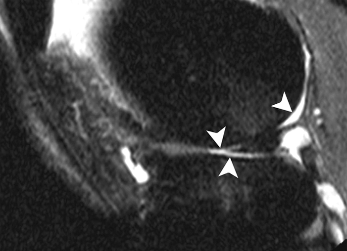
Figure 1b:

Sagittal intermediate-weighted fat-suppressed MR images (4800/35) show fast cartilage loss in lateral compartment between (a) baseline and (b) follow-up. (a) Maceration (arrowhead) of anterior horn of lateral meniscus. (b) Diffuse cartilage loss in central and posterior parts of lateral femur and in central region of lateral tibia (arrowheads).
