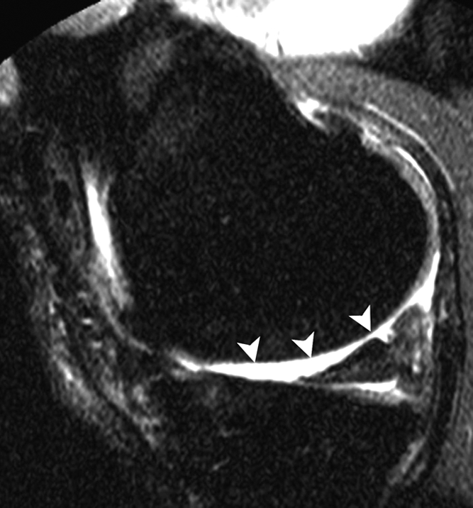
Figure 2c:

Sagittal intermediate-weighted fat-suppressed MR images (4800/35) show fast cartilage loss in medial compartment between (a,b) baseline and (c) follow-up. (a) Effusion (black arrow) and marked synovitic infiltration of Hoffa fat pad in intercondylar (arrowheads) and infrapatellar (white arrow) regions. (b) Small superficial cartilage defect (arrow) in central region of medial femoral condyle. (c) Massive cartilage loss and denudation of bone (arrowheads) in central region of medial femoral condyle. Note also diffuse cartilage damage in central part of tibial plateau.
