
Figure 2b:
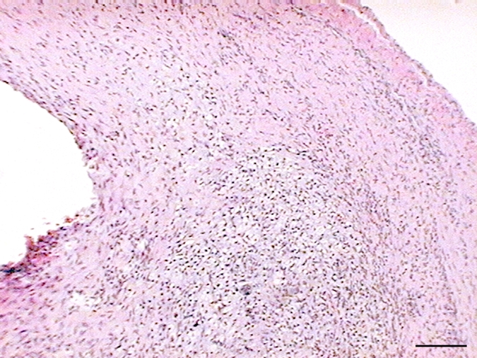
(a–f) Examples of histologic findings. (Original magnification, ×10.) Hematoxylin-eosin–stained (a) control aneurysm and (b) LPS-injected aneurysm. Anti–MPO-antibody staining is (c) absent in control aneurysms and (d) strongly positive in LPS-injected aneurysms. MAC387 staining was (e) absent in control aneurysms and (f) strongly positive in LPS-injected aneurysms. (g) Bar graph shows significantly (**) increased MPO activity in the LPS-injected aneurysms compared with control aneurysms and left CCAs (P < .002). Bars represent 0.1 mm.