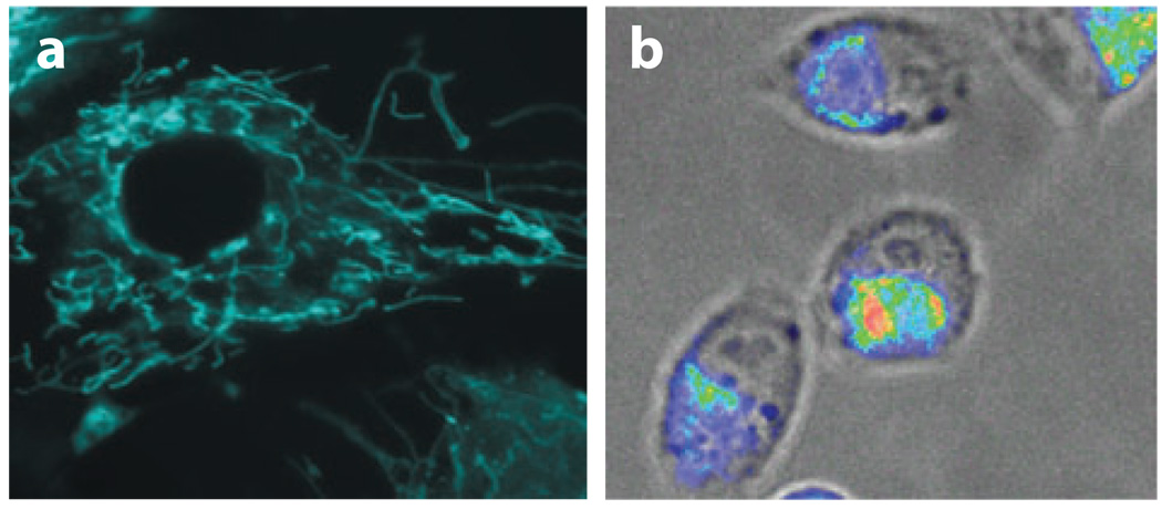
Figure 6.
Imaging mRNA localization in HDF and MIAPaCa-2 cells using dual FRET molecular beacons targeting K-ras and survivin mRNA, respectively. (a) Fluorescence images of K-ras mRNA in stimulated HDF cells. Note the filamentous K-ras mRNA localization pattern. (b) A fluorescence image of survivin mRNA localization in MIAPaCa-2 cells. Note that survivin mRNAs often localized to one side of the nucleus of the MIAPaCa-2 cells.

