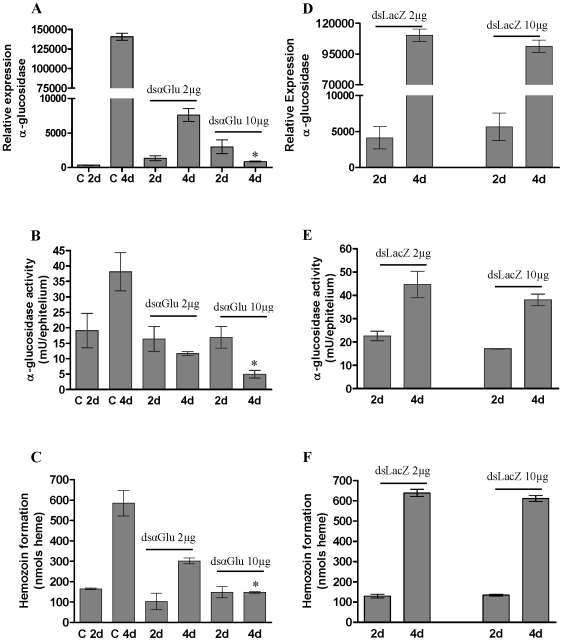
Figure 3. Silencing of α-glucosidase by injection of dsRNA and its action in R. prolixus “in vivo”.
A: Relative expression (qPCR) of α-glucosidase in the midgut after injection of dsαglu; B: α-glucosidase activity into midgut of insects injected with dsαglu; C: Hz produced by the insects injected with dsαglu; D: Relative expression of α-glucosidase (qPCR) in the midgut after injection of dsLacZ; E: α-glucosidase activity into midgut of insects injected with dsLacZ; F: Hz produced by the insects injected with dsLacZ. The insects were injected with 2 µL of 100 mM PBS pH 7.4 (control), dsLacZ (2 or 10 µg/female) or dsα-glu (2 or 10 µg/female) and analyzed 2 or 4 days after feeding on blood. Hz measurement was carried out using a pool of six midgut epithelium. Four replicates were performed. Each replicate consists of a pool of six adult females. The insects injected with 10 µg dsRNA after 4 days of feeding were significantly different from control 4 day insects *(P<0.05).

