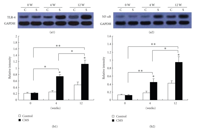
Figure 6.
Validation of changes in TLR-4 (73 KD) and NF-κB p65 (80 kDa) protein expression by western blotting. Proteins extracted were prepared from arteries (n = 6) of mice subjected to CMS or normal conditions for 0, 4, and 12 weeks, respectively. Each lane shows representative western blots using anti-TLR4 or NF-κB p65 and anti-GAPDH bodies in (a1) and (a2), respectively. Each panel summarizes densitometric readings of band intensities normalized to GAPDH, which was measured by densitometry with Image J image analysis software. (b1), (b2) densitometric measurements TLR4 and NF-κB p65 from Western blots, respectively. Data are mean ± SEM. C: control group; S: chronic mild stress group (n = 6 per group). *P < .05, **P < .01 compared with the control. The data are representative of three experiments.

