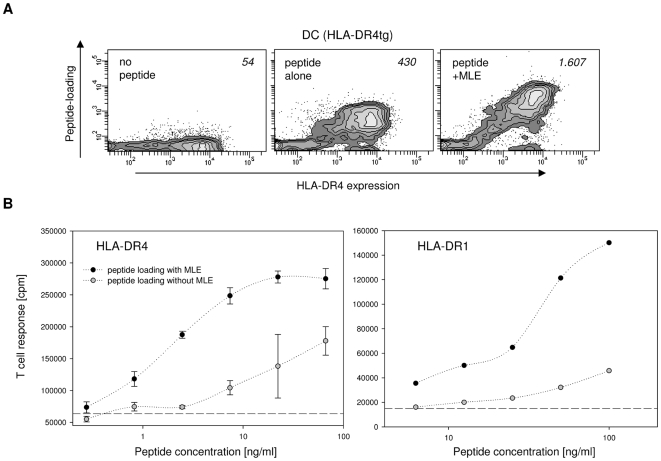
Figure 1. Influence of MLE on the class II MHC peptide-loading of dendritic cells.
(A) Cell surface loading. HLA-DR4 expressing dendritic cells (DC) generated from the bone marrow of HLA-DR4 transgenic mice were incubated for 4 h with medium alone (left panel) or with 5 µg/ml biotinylated HA 306–318 peptide in the absence (middle panel) or presence of 250 µM AdEtOH, the model MLE compound used throughout this study (right panel). Contour plots are shown for DC after staining with anti-HLA-DR antibody (→ MHC expression) and streptavidin (→ peptide load). Mean peptide loading (MFI of streptavidin signal) is indicated. (B) CD4+ T cell response. DC from HLA-DR4tg mice (left panel) and from HLA-DR1tg mice (right panel) were pulsed for 4 h with indicated amounts of HA 306–318 peptide in the absence (open circle) and presence (closed circle) of 250 µM AdEtOH. The cells were used to challenge HA 306–318 specific, HLA-DR4-restricted 8475/94 cells and HLA-DR1-restricted EvHA/X5 T cell hybridoma cells, respectively. Background proliferation was measured in absence of peptide (dashed line).

