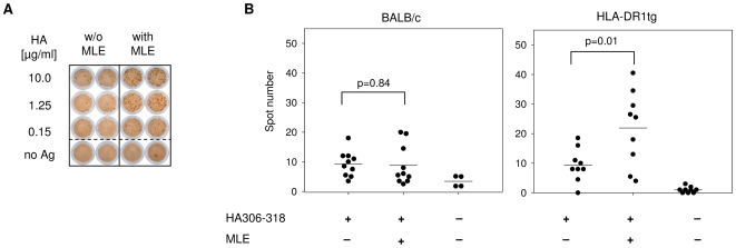
Figure 3. Vaccination in presence of MLE increases the number of antigen-specific IFNγ-producing T cells.
(A) Determination of IFNγ in an Elispot assay. 12 days after vaccination, 1×10⁶ lymph node cells from mice primed with 3 µg HA 306–318 in IFA/CpG supplemented without (left panel) or with AdEtOH (right panel) were incubated with titrated amounts of HA306–318 peptide on a plate coated with α-IFNγ antibody. Detection was carried out 48 hrs later by determining the spot number in each well. Spots represent single IFNγ+ cells. (B) Statistical analysis of the T cell response. Summarized Elispot data obtained from groups of BALB/c mice (left panel, n = 10) and HLA-DR1tg mice (right panel, n = 9) were analyzed using student's t test.

