
Figure 7.
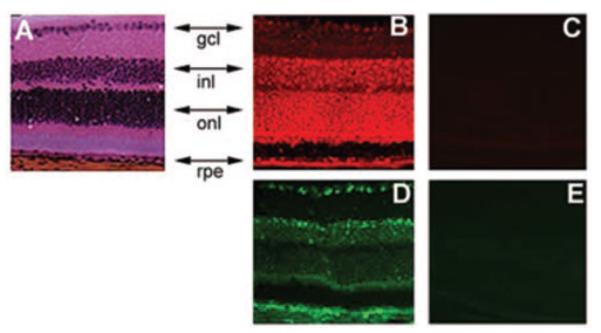
Immunolocalization of the two subunits of system xc- (xCT and 4F2hc) in mouse retina. Retinal sections were probed with antibodies specific for each of these subunits. (A) Hematoxylin and eosin staining. (B) Expression pattern of xCT as detected by Cy-3–conjugated secondary antibody (red fluorescence). (C) Negative control for xCT expression where the primary antibody was first neutralized with excess antigenic peptide and then used for incubation with retinal sections. (D) Expression pattern of 4F2hc as detected by FITC-conjugated secondary antibody (green fluorescence). (E) Negative control experiment for 4F2hc expression, in which the primary antibody was omitted. rpe, retinal pigment epithelium; onl, outer nuclear layer consisting of photoreceptor cells; inl, inner nuclear layer consisting of Müller cells, and neuronal cells (bipolar, amacrine, and horizontal cells); gcl, ganglion cell layer.