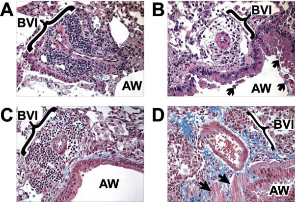
Figure 7.
Increased bronchovascular collagen deposition is evident during later stages of C. neoformans infection in CCR2−/− mice. CCR2+/+ (A, C) and CCR2−/− (B, D) mice were inoculated IT with C. neoformans and lungs were harvested at day 21 post-infection. Samples were processed for histological evaluation as described in Methods. (A), Photomicrographs (H & E staining, ×400 magnification) in CCR2+/+ (A) and CCR2−/− (B) mice depict a persistent reduction in bronchovascular infiltrates (BVI) adjacent to airways (AW) in CCR2−/− mice. Trichrome staining in CCR2+/+ (C) and CCR2−/− (D) mice (×400) reveals increased bronchovascular fibrosis (blue) in CCR2−/− mice. Note that airway mucus production (B, arrows) and smooth muscle (D, block arrowheads) are more apparent in CCR2−/− mice.

