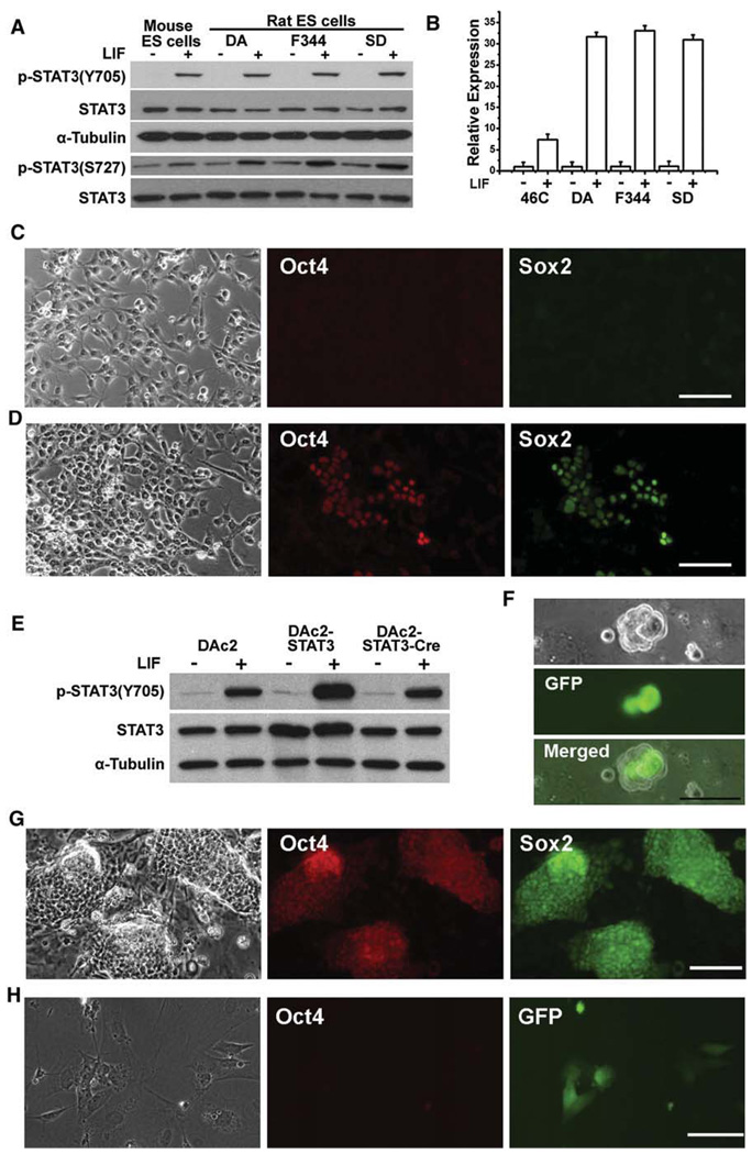
Figure 6. Rat ES Cells Are Responsive to LIF/STAT3 Signaling for Self-Renewal.
(A) Analysis of STAT3 activation by western blot in mouse and rat ES cells stimulated with 10 ng/ml LIF for 30 min.
(B) qRT-PCR analysis of Socs3 induction by LIF treatment in mouse 46C ES cells and rat ES cells. Data represent mean ± SD of triplicate samples from two independent experiments.
(C and D) Immunofluorescence staining for Oct4 and Sox2 in DAc2 rat ES cells 7 days after they were cultured in laminin/N2B27 (C) or laminin/N2B27+LIF (D) conditions. Scale bars represent 50 µm.
(E) Western blot analysis of STAT3 activation in DAc2, DAc2-STAT3, and DAc2-STAT3-Cre rat ES cells after treatment with 10 ng/ml LIF for 30 min.
(F) DAc2-STAT3 cells, 1 day after transient transfection with Cre to remove the STAT3 transgene and simultaneously activate GFP. The scale bar represents 50 µm.
(G) Immunofluorescence staining for Oct4 and Sox2 in DAc2-STAT3 cells after nine passages in L cell/LIF conditions. The scale bar represents 50 µm.
(H) Immunofluorescence staining for Oct4 in DAc2-STAT3-Cre cells at second passage in L cell/LIF conditions. The presence of GFP denotes the removal of the STAT3 transgene. The scale bar represents 50 µm.

