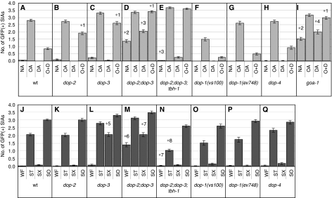
Figure 3.
CRE-mediated GFP expression of dopamine receptor and G protein mutants. (A–I) The number of GFP-expressing SIA neurons per animal was determined after animals were incubated on food containing plates also containing no amine (NA), octopamine (OA), dopamine (DA), or octopamine+dopamine (O+D) for 6 h. (J–Q) The number of GFP-expressing SIA neurons per animal was determined after animals were incubated in well-fed (WF), starvation (ST), starvation+Sephadex (SX), or soaking (SO) conditions for 6 h. The data in panels A and J are also shown in Figures 1E and 2K, respectively, and are shown here for comparison. Error bars indicate the standard errors of the means. At least 80 animals were tested. *1P<0.001 (Tukey–Kramer multiple comparison test), compared with O+D of wt animals. *2P<0.001, compared with NA of wt animals. *3P<0.001, compared with WF of dop-2;dop-3 double mutants. *4P<0.01, compared with WF of goa-1 mutants. *5P<0.001, compared with SX of wt animals. *6P<0.001, compared with WF of wt animals. *7P<0.01, compared with WF of dop-2;dop-3 double mutants. *8P<0.001, compared with WF of dop-2;dop-3;tbh-1 triple mutants.

