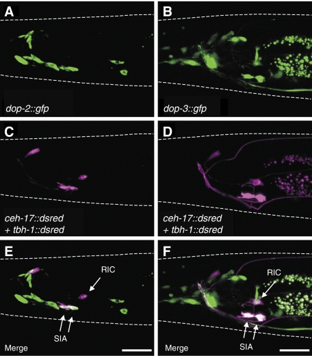
Figure 4.
Expression patterns of dop-2 and dop-3. Fluorescence images for (A, B) GFP expression and (C, D) DsRed expression were obtained from animals carrying ceh-17∷dsred and tbh-1∷dsred in addition to (A, C) dop-2∷gfp or (B, D) dop-3∷gfp. (E, F) Merged images of (A+C) and (B+D), respectively. dop-2∷gfp induced GFP expression in the SIA neurons but not in the RIC neurons. dop-3∷gfp induced GFP expression in the SIA neurons and the RIC neurons. Both dop-2∷gfp and dop-3∷gfp also each induced GFP expression in some other cells as reported earlier (Suo et al, 2003; Tsalik et al, 2003; Chase et al, 2004). White dotted lines outline the head of each animal. Scale bars, 20 μm.

