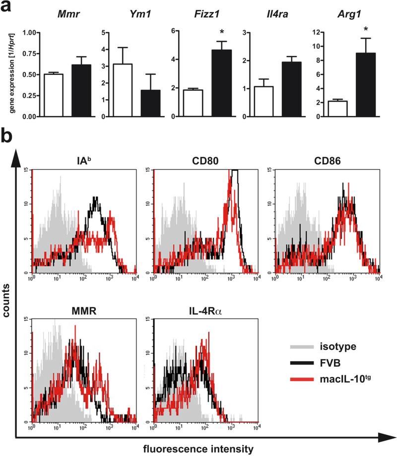
Figure 7. Enhanced alternative macrophage activation in lungs from Mtb-infected macIL-10tg mice.
FVB (open symbols) and macIL-10tg (solid symbols) mice were infected with 100 CFU Mtb via the aerosol route. (a) Gene-expression of Mmr, Ym1, Fizz1, Il4ra, Arg1 was determined in lung homogenates from mice infected for 42 days by quantitative real time RT-PCR based on expression of hprt. Statistical analysis was performed using the unpaired Student's t test defining differences between FVB and macIL-10tg mice as significant (*, p≤0.05). (b) Expression of activation markers on pulmonary macrophages was assessed by flowcytometric analysis of IAq, CD80, CD86, MMR, and IL-4Rα gated on PI− F4/80+ cells in single cell suspensions of perfused lungs from FVB (black line) and macIL-10tg mice (red line) mice. Representative histogram of 1 out of 3 mice per group (gray histogram, isotype control).

