Figure 7.
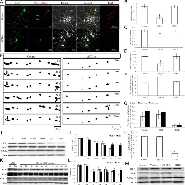
Effects of ADDLs on mitochondrial morphology and distribution in primary neurons. Primary rat E18 hippocampal neurons (DIV 9–12) transfected with GFP and Mito-DsRed2 were treated with 800 nm ADDLs for 24 h. Twenty-four hours of treatment of Aβ42-1, subject to the same procedure that produces ADDLs, was used as a control. A, Representative pictures of positively transfected neurons. Red, DsRed; green, GFP; white: tubulin staining. B–E, Quantification of mitochondrial length (B), density (C), neurite mitochondrial index (D), and axial length of neurites devoid of mitochondria (E) in a segment of neuronal process 400 μm in length beginning from the cell body of neurons (*p < 0.05, Student's t test). At least 20 cells were analyzed in each experiment, experiments were repeated three times. F, Demonstration of the effect of ADDLs on mitochondrial fission and fusion events. Rat E18 hippocampal neurons (DIV 9) were transfected with Mito-DsRed2. Twenty-four hours after incubation with or without 800 nm ADDLs at DIV 11, neurons were imaged in time lapse (10 s interval, 20 min). Representative thresholded time-lapse pictures showed active mitochondrial fission and fusion in the segment of axon ∼100 μm in length beginning 300 μm from the cell body of control or ADDL-treated neurons. Active mitochondrial fission (filled arrows) and fusion (empty arrows) and fast-moving mitochondria (asterisks) are marked. G, H, Both fusion and fission were impaired significantly by ADDLs (G), and mitochondria spent significantly less time in the post-fusion fused state than in the post-fission single state (H). At least 20 neurons were analyzed in three independent experiments (*p < 0.05, Student's t test). I–L, Immunoblot and quantitative analysis of DLP1 and OPA1 levels in neurons treated at the indicated dosages of ADDLs for 24 h (I, J) or at 800 nm for the indicated periods of time (K, L) revealed that ADDLs induced a dose- and time-dependent decrease in DLP1 and OPA1 levels (*p < 0 0.05, Student's t test). C, Control, UNT, untreated. M, Unlike ADDLs, 10 μm Aβ42-1 had no effect on the expression of DLP1 and OPA1. Equal protein amounts (15 μg) were loaded. Actin immunoblot was used as an internal loading control.

