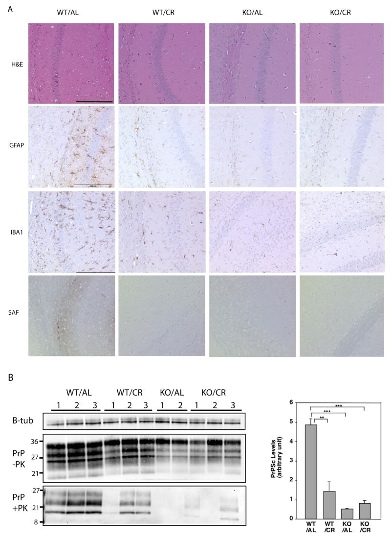
Figure 1.
The onset of prion disease is delayed in calorie restricted mice and SIRT1 knockout mice. Wild type (WT) mice and SIRT1 KO mice fed AL or on CR diets were injected with RML prions. Brains were removed 4 months post inoculation. (A) Brain sections were stained with hematoxylin and eosin to visualize vacuolation, anti-GFAP to visualize gliosis, anti-IBA1 staining to visualize microglia, and anti-PrP (SAF) on formic acid treated samples to visualize aggregates of PrP. Scale bars correspond to 200um. (B) Brain homogenates were subjected to proteinase-K digestion. Proteinase resistant PrP was detected by western blotting with anti-PrP antibody. β-tubulin (from undigested homogenates) was used as a loading control. The level of PK-resistant PrP was quantitated using ImageJ software.

