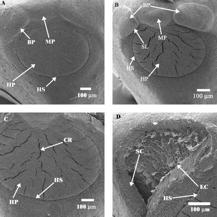
Fig. 5.

Electron micrographs of non-treated (dormant) and of treated (non-dormant) seeds: (A) hilum–microphyle area of a dormant seed; (B) hilum–micropyle area of a non-dormant seed; (C) hilum pad of a non-dormant seed; (D) opening where bulge has become detached from seed coat. BP, Bulge; CR, crack; EC, endodermal cells; HP, hilum pad; HS, hilar fissure; MP, micropyle; SC, seed coat showing palisade cells and sclereids; SL, slit through which water enters the seed.
