
Fig. 2.
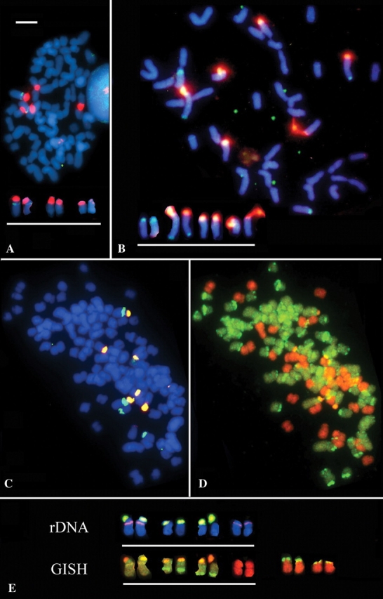
(A–C) FISH probed with 18–26S rDNA (red or orange) and 5S rDNA (green) and chromosomes counterstained with DAPI (blue) in (A) metaphase cell of I. virginica showing three loci (six signals at diploid) of 18–26S rDNA signals and one locus of 5S rDNA on one of the 18–26S carrying chromosomes (labelled chromosomes are shown arranged as a karyotype below metaphase); (B) metaphase of I. setosa with three 18–26S rDNA loci and two pairs of 5S rDNA loci of different sizes; the smallest 5S locus occurs on the same chromosome as a 18–26S rDNA locus. (C) metaphase cell of I. versicolor showing six signals of 18–26S rDNA and four signals of 5S rDNA. (D) The same cell reprobed by GISH using total genomic DNA from I. virginica (green) and from I. setosa (orange). (E) Chromosomes underlined are isolated rDNA-carrying chromosomes from an I. versicolor metaphase labelled with 18S–26S (yellow–green) and 5S (pink) rDNA, and the same chromosomes after GISH labelled in (D). From the same metaphase the two pairs of chromosomes to the right are predominantly labeled with I. setosa genomic DNA but show translocations from I. virginica-origin chromosomes. Scale bar of complete metaphases represents 10 µm.