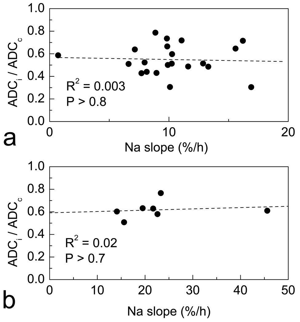
Figure 3.
23Na slope (the rate of [Na+]br increase) and ADC deficit (ADC ratio of ipsilateral to homotopic contralateral ROIs, ADCi/ADCc) in the same brain ROIs in ischemia. (a) In the typical brain (rat #6), ADC deficit shows no correlation with slope in ROIs characterized as ischemic by ADC (i.e., ADCi/ADCc < 0.8). (b) ADC deficit in the ROIs of maximum slope of different rats shows no correlation with the maximum slope values. Each data point corresponds to an individual animal for which both parameters were available (n = 7).

