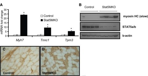
Figure 4.
Type I fiber gene expression in quadriceps muscle fibers from Stat5MKO and control mice. A) Real-time PCR gene expression analysis of selected slow-isoform genes (n=3). B) Western blot analysis of 4 control and 4 Stat5MKO animals probed for slow-twitch myosin heavy chain (Myh7, top panel), STAT5a/b (middle panel) and b-actin (bottom panel) to show loading consistency. C, D) Control and Stat5MKO quadriceps muscles from 14-wk-old animals from each group were analyzed. Transverse sections were stained for myosin heavy chain slow isoform. C) Type I myosin staining in quadriceps muscle of control mice. D) Type I myosin staining in quadriceps muscle of Stat5MKO shows mostly fast-twitch; however, tissue contains areas of abundant type I fibers not seen in controls. View shows area of abundant type I fibers.

