Figure 1.
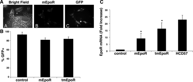
Lentiviral transduction of EpoR in myoblasts. A) Immunostaining of EpoR expression and GFP fluorescence in 293T cells after transduction of LV-mEpoR-GFP for 24 h. B) GFP-positive myoblasts were detected by fluorescence microscopy after lentiviral infection for 48 h and counted as a percentage of GFP-positive staining cells. C) Mouse EpoR mRNA expression in myoblasts after lentivirus infection by quantitative real-time RT-PCR. C2C12 cells treated with LV-mEpoR-GFP and LV-tmEpoR-GFP were harvested 48 h after lentivirus infection. Untreated C2C12 cells were used as negative control; and HCD57 cells were used as positive control. Mouse S16 mRNA expression was used as internal control. Mouse EpoR mRNA expression levels significantly increased in LV-mEpoR-GFP and LV-tmEpoR-GFP-infected C2C12 cells compared to control cells treated with LV-GFP. *P < 0.01.

