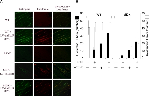
Figure 6.
tmEpoR overexpression in grafted myoblasts favors formation of functional muscle fibers in mdx mice. C2C12 cells treated with LV-tmEpoR-Luc or LV-GFP-Luc control were injected at 2-mm depth into the gastrocnemius muscle of 3-mo-old mdx and WT mice. Erythropoietin (3000 U/kg) was administered by intraperitoneal injection to the EPO treatment groups on the same day of myoblast transplantation. After 6 wk of myoblast transplantation, mice were anesthetized and perfused with 4% paraformaldehyde in PBS. Gastrocnemius muscle was collected and sectioned for immunohistochemistry luciferase (red) and dystrophin (green) staining. A) Representative pictures of gastrocnemius muscle sections stained for dystrophin (left column), luciferase (center column), and an overlap of dystrophin/luciferase (right column) in WT and mdx mice grafted with LV-tmEpoR-Luc C2C12 cells and exposed to EPO. B) Quantification of luciferase and dystrophin-positive fibers in WT and mdx mice (positive-stained fibers/20 random field views at ×40).

