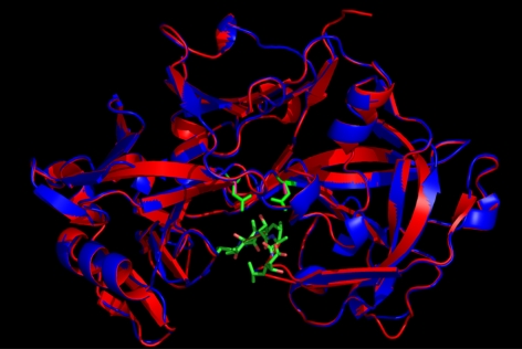
Figure 3.
Molecular model of Na-APR-1wt (red ribbon with green active-site Asp side chains) and Na-APR-1mut (red ribbon with fuschia active-site Ala side chains) superimposed on the template structure of human pepsin (blue ribbon) (PDB code 1PSO). Structure of 1PSO was solved in complex with pepstatin, shown in stick representation with carbon atoms in green, nitrogen atoms in blue, and oxygen atoms in pink. RMSD over the c-α atoms of the model and the template is 0.118, highlighting the overall similarity between the two molecules. Superimposition was produced using the program VMD (43).

