Figure 4.
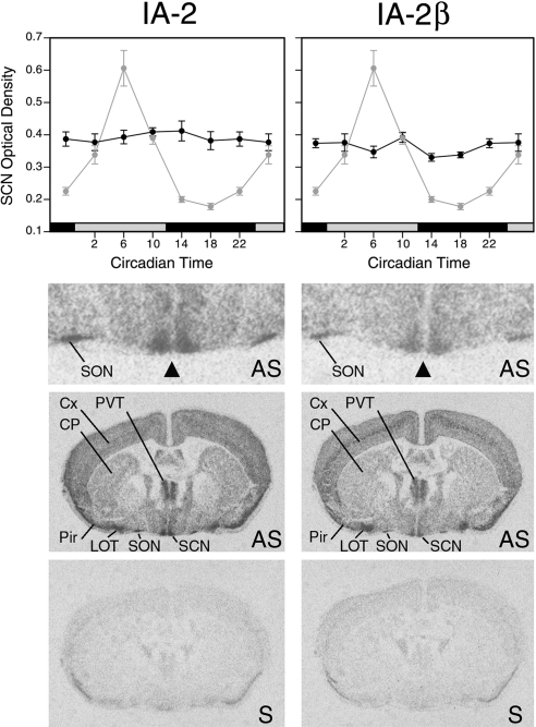
Expression of IA-2 (left) and IA-2β (right) (dark black lines) as determined by in situ hybridization in brain sections through the SCN from WT mice housed in constant darkness (top panels). IA-2 and IA-2β showed no evidence of rhythmicity (P>0.05; ANOVA,). Adjacent sections from the same animals revealed the expected high-amplitude rhythm of mPer1 expression (light gray symbols with same line plotted in both left and right panels). Values represent means ± se of 5 to 6 animals. Sections hybridized with antisense (AS) probes showed that both IA-2 and IA-2β genes are highly expressed in the SCN, with no obvious subregional localization. Top image is a higher magnification of the image below it. Triangle indicates the bilaterally symmetrical SCN. Sense (S) strand control probes showed the level of the background hybridization (bottom panels). Cx, cortex; CP, caudate-putamen; LOT, nucleus of the lateral olfactory tract; Pir, piriform cortex; SON, supraoptic nucleus; PVT, paraventricular nucleus of the thalamus.

