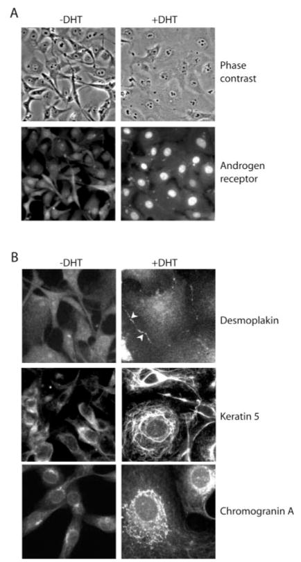
Fig. 2. DHT treatment of HT-AR1 cells induces cytoskeletal reorganization and neuroendocrine-like differentiation.
A, cell morphology and AR protein expression in HT-AR1 cells cultured for 3 days on fibronectin-coated coverslips in Dulbecco’s modified Eagle’s media containing 5% C/S CBS serum with or without 10 nM DHT. AR protein was detected by immunostaining using an anti-AR antibody. The same field of cells is shown by phase-contrast or fluorescence microscopy. B, HT-AR1 cells cultured the same as in A and stained with antibodies that specifically recognize desmoplakin, keratin 5, or chromogranin A. The cells in the keratin 5 immunostaining were counter-stained with Hoechst dye to visualize the nuclei.

