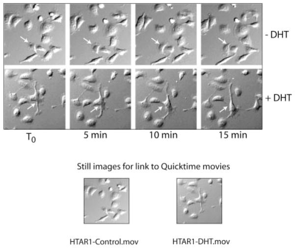
Fig. 3. Time lapse video microscopy of HT-AR1 cells shows rapid changes in membrane morphology in DHT cultures.
Cells were seeded on a glass Delta T dish and grown overnight in Dulbecco’s modified Eagle’s selection media and then changed to culture media the next day with or without DHT. Images were captured every 5 min beginning with T0, which was 1 h after adding culture media ±DHT. Representative fields from the two cultures are shown of the first 15 min of video microscopy. The arrows identify individual cells in the same field over the time course. QuickTime videos of these two cultures out to 5 h are included as Supplemental Materials (available at http://www.jbc.org).

