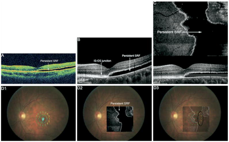
Figure 3.
A, Stratus OCT of Patient 2 showing shallow SRF involving the fovea 12 months after pneumatic retinopexy. Visual acuity was 20/20, and the patient had no subjective visual symptoms. B, FD OCT B-scan also shows persistent SRF and an unremarkable photoreceptor layer (IS-OS junction) in the detached and attached macula. C, Flattened FD OCT C-scan indicating the extent of SRF in the macula. D1, MP with threshold values shown. Small, light blue dots indicate the patient’s fixation. D2, Flattened C-scan overlain onto fundus photograph. D3, Flattened C-scan superimposed onto MP indicates decreased sensitivity (yellow dots) in the temporal area of persistent SRF (black circle). SRF = subretinal fluid; IS-OS = inner segment-outer segment.

