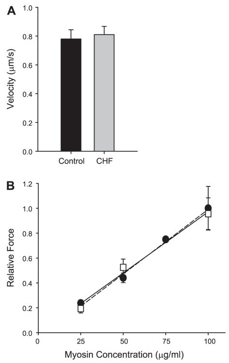
Fig. 4.
A: the velocity of actin filaments propelled by myosin isolated from the vastus lateralis muscle of CHF and control research subjects. Actin is isolated from chicken pectoralis. B: relative isometric force as a function of skeletal myosin concentration in the loading buffers for CHF (squares, solid regression) and control (circles, dashed regression) research subjects. Force increased as a function of myosin concentration with no difference demonstrated between the two groups.

