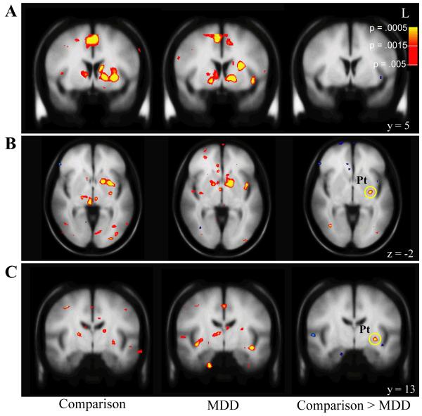
FIGURE 2.
Reward-related anticipatory activation in MDD (N=26) and comparison (N=31) subjects.
Coronal (A) and axial (B) slices showing anticipatory reward activity [Reward cue - No-incentive cue] in basal ganglia regions are shown for both comparison and MDD subjects, as well as for the random effect analyses comparing the two groups. (A) Robust activation of ventral striatal regions, including the nucleus accumbens, is seen in both groups, leading to a lack of group differences. (B) Relative to comparison subjects, the MDD group shows significantly reduced activation during reward anticipation in the left putamen (x=-28, y=-13, z=-2). All contrasts are thresholded at p<0.005. Pt = Putamen, L = Left

