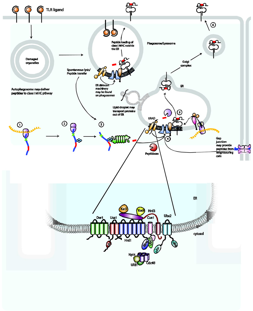
Figure 3. Additional pathways that may be relevant for antigen processing and presentation by MHC class I molecules in TLR-stimulated DCs.
How peptides traffic of peptides to MHC class I molecules during cross-presentation in DCs is unknown. We propose several possibilities that could permit peptides to access MHC class I molecules. Autophagosomes containing both endogenous peptides and pathogen-derived proteins could potentially serve as a source of peptides for the MHC class I pathway. Peptides may transit from the phagosome through leaky membranes, following spontaneous lysis of the phagosomal compartment or by traversing the lipid bilayer. Components of the peptide-loading complex and the endoplasmic reticulum (ER) dislocation machinery may be shuttled from the ER to the phagosome, perhaps involving lipid droplets for transport. Transfer of these components to the phagosome may permit loading of MHC class I molecules with peptides at this site rather than in the ER. Gap junctions may serve as conduits to allow neighbouring cells to donate peptide epitopes for loading of MHC class I molecules. The yeast ERAD protein complex is illustrated in greater detail on the panel below.

