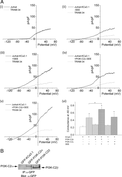
Figure 2.
TCR stimulation of Jurkat T-cells activates KCa3.1 channel activity. Jurkat T-cells overexpressing KCa3.1 (Jurkat-KCa3.1) were transfected with GFP, GFP-PI3K-C2β, or GFP-PI3K-C2α and incubated with Raji B-cells that were either untreated or treated with the superantigen SEE as described in Materials and Methods. (A, ii–v) Whole-cell patch- clamp was then performed on GFP-positive cells that were incubated with Raji B-cells that were either untreated or treated with the superantigen SEE as described in Materials and Methods. (vi) Bar graph summary of KCa3.1 conductance (pS) measured at −60 mV from 10 independent experiments. (A, i) shows control Jurkat T-cells do not have KCa3.1 channel activity. *p < 0.05 compared with KCa3.1 conductance measured at −60 mV without the addition of SEE in each group and as indicated in the figure. (B) α-GFP Western blot of GFP-sorted cells demonstrating equal protein expression of GFP-PI3K-C2α and PI3K-C2β.

