Figure 3.
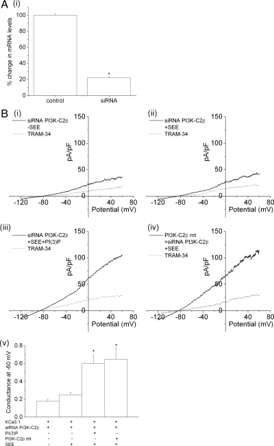
siRNA knockdown of GFP-PI3K-C2β inhibits TCR-stimulated activation of KCa3.1 channel activity. (A) Real-time PCR showing >80% silencing of PI3K-C2β in siRNA PI3K-C2β siRNA-transfected cells. *p < 0.05 compared with control. (B) Jurkat-KCa3.1 cells were transfected with a pool of siRNAs to PI3K-C2β and stimulated 48 h after transfection with Raji B-cells that were either untreated (i) or treated with SEE (ii). Whole-cell patch clamp was then performed as described in Figure 2. (iii) To determine whether inhibition of KCa3.1 channel activity in siRNA PI3K-C2β–transfected cells was due to decreased levels of PI(3)P, siRNA PI3K-C2β–transfected cells were dialyzed with PI(3)P as described in Figure 1. (iv) In addition, KCa3.1 channel activity was rescued by transfecting a GFP-PI3K-C2β mutant (PI3K-C2β mt) that abrogated interaction with the siRNA. (v) Bar graph summary of KCa3.1 conductance (pS) measured at −60 mV (n = 10 cells). *p < 0.05 compared with siRNA PI3KC2β + SEE–transfected cells.

