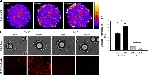
Figure 1.
Insulin induces cortical actin remodeling in adipocytes as shown by TIRFM. Actin-eGFP expressing 3T3-L1 adipocytes were serum-starved for 120 min before imaging by TIRFM at 10 Hz for 2 min before stimulation with 100 nM insulin (at t = 0) and then were imaged for a further 20 min. (A) Representative images are shown from the time course. (B) 3T3-L1 adipocytes were serum-starved for 120 min and treated with DMSO ± 10 μM Lat-B for 1 h. Cells were then either unstimulated (Basal) or stimulated with 100 nM insulin (Insulin) for 30 min at 37°C, fixed, and labeled with TRITC-phalloidin then imaged by TIRFM. Representative images are shown. (C) The mean fluorescence of cells from three separate experiments are shown; # p < 0.05. Scale bar, (A and B) 10 μm.

