Figure 7.
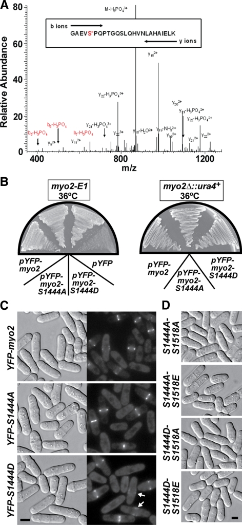
Cytokinesis is not dependent on the phosphorylation state of the Myo2p tail at Ser-1444 or -1518. (A) Electrospray ionization liquid chromatography ion trap mass spectrometry was performed on the Myo2p heavy chain. The Coomassie-stained heavy-chain band was cut out of an SDS-PAGE gel and analyzed. The phosphorylated peptide GAEVS*PQPTGQSLQHVNLAHAIELK produced a precursor ion at m/z 902.35 (z = 3) and was selected for MS/MS fragmentation. The largest MS/MS ion, m/z 869.93 (z = 3) corresponded to the precursor ion with the neutral loss of phosphate (−97.97 Da). The second and third largest MS/MS ions m/z 978.84 and 1091.83 (z = 2), corresponded to fragment ions y18 and y20, respectively. These ions are generated from cleavage of the precursor ion on the N-terminal side of Pro1445 or Pro1447, respectively, and their high abundance is anticipated (Breci et al., 2003). The MS/MS ions m/z 426.09, 523.22, and 651.21 (z = 1) correspond to fragment ions b5, b6, and b7 with the neutral loss of phosphate. These fragment ions indicate phosphorylation is located on Ser-1444 rather than Ser-1451. (B) pYFP-myo2-S1444A and -S1444D complement the temperature-sensitive lethality of the myo2-E1 mutant (left plate) and rescue growth of a myo2Δ strain (right plate). pYFP-myo2: positive control; pYFP: negative control lacking a myo2 insert. Cells were streaked out and grown at 36°C on EMM minimal media plates lacking leucine. (C) Gene replacement of myo2 with YFP-myo2-S1444A or -S1444D forms does not impact cytokinesis (left panels, DIC). Localization of integrated YFP-Myo2p, YFP-Myo2p-S1444A, and YFP-Myo2p-S1444D was captured by epifluorescence microscopy (right panels). Arrows in the bottom right panel highlight the presence of YFP-Myo2p-S1444D in the broad bands of assembling contractile rings. Bar, 4 μm. (D) Morphologies of YFP-myo2-S1444A-S1518A, YFP-myo2-S1444A-S1518E, YFP-myo2-S1444D-S1518A, and YFP-myo2-S1444D-S1518E double mutants. All cells were grown at 32°C in YE5S media before imaging. Bar, 4 μm.

