
Figure 1. C2GlcNAcT-I deficiency suppresses injury-induced arterial neointima formation.
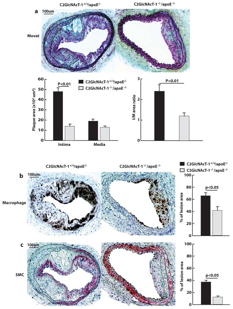
a, Cross-sections of arterial neointima stained with Movat pentachrome and quantification of the size of neointima (I), size of media (M), and ratio of intima to media (n = 12 for both groups). b, Anti-Mac-2 staining of infiltrated macrophages in arterial neointima. c, Anti-α-actin staining of vascular smooth muscle cells (SMCs) in the arterial neointima. The average numbers of cells in the neointima were obtained by analyzing 12 cross sections from 12 injured carotid arteries from mice.