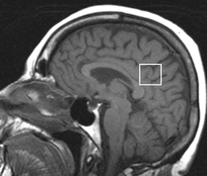
Figure 1:
Location of 1H MR spectroscopy voxel. The 1H MR spectroscopy voxel was placed on a midsagittal T1-weighted localizer image (700/14). The anterior border of the splenium, superior border of the corpus callosum, and cingulate sulcus were the anatomic landmarks used to define the anteroinferior and anterosuperior borders of the 8-cm3 voxel. This voxel partially included the right and left posterior cingulate gyri and the inferior precuneate gyri (portions of Brodmann areas 23 and 31) in both hemispheres.

