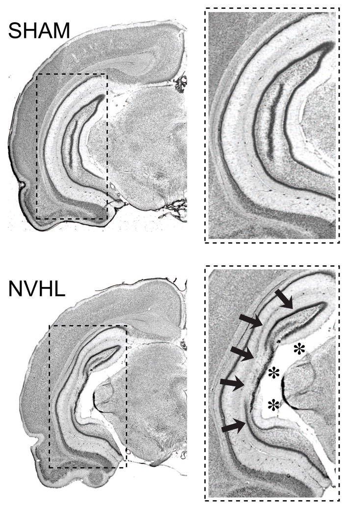
Figure 4.
Photomicrographs depicting the extent of a typical neonatal ventral hippocampal lesion. Coronal Nissl stained sections show the ventral hippocampus of a sham-operated rat (top, SHAM) and a characteristic neonatal ventral hippocampal lesion (bottom, NVHL), characterized by cell loss (arrows), and enlarged ventricles (asterisks).

