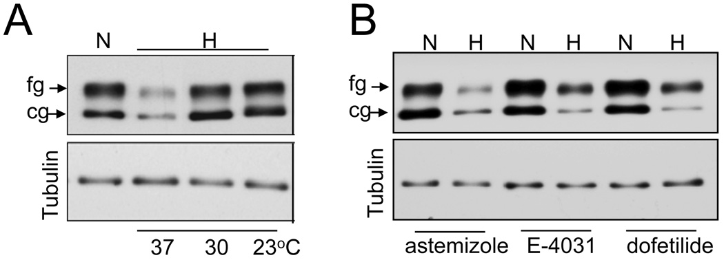
Fig 1. Effect of temperature and antiarrhythmic drugs on hERG expression during hypoxic exposure.
A. Immunoblot analysis of HEK293/hERG cell lysates exposed to 24hrs of normoxia (N), and hypoxia (H) at 37°C, 30°C, 23°C. B) Cells treated with antiarryhthmic drugs astemizole (5µM), dofitilde (3µM) and E-4031 (5µM) were exposed to hypoxia at 37°C for 24 hrs and cell lysates analyzed for hERG expression by immnoblots. Tubulin protein expression was used as a loading control.

