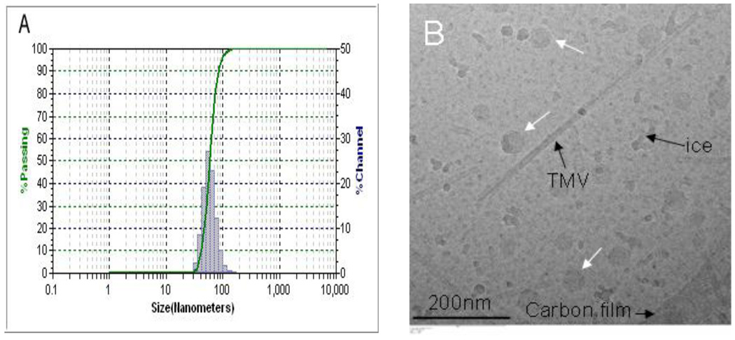
Fig. 3.
The size distribution of PEG5k-CA8 nanoparticles loaded with PTX and NIRF dye DiD in PBS measured by dynamic light scanning (DLS) (A) and Cryo-Transmission electron microscopy (Cryo-TEM) (B). In Fig. 3B, white arrows point to nanoparticles, while Tubacco Mosaic Virus (TMV) was used as calibration standard (18 nm in width).

