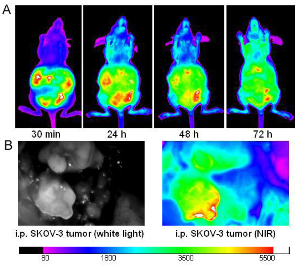
Fig. 6.
Intra-abdominal distribution of PEG5k-CA8 nanoparticles. (A) In vivo NIRF imaging of the intraperitoneal SKOV-3 tumor bearing mice at different time points after i.p. injection of DiD-PTX-NPs. (B) Localization of DiD-PTX-NPs on tumors. The mice were sacrificed at 72 h post injection, and the abdominal cavity was exposed to scan with Kodak imaging station.

