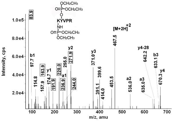
Figure 7.
A CID mass spectrum of the diethoxyphosphate-labeled, bovine tubulin beta, tryptic peptide K*Y*VPR. The values enclosed in the boxes are the masses of the characteristic fragments for diethoxyphosphate-labeled lysine and diethoxyphosphate-labeled tyrosine. The parent ion is marked by [M+2H]+2. Lysine 58 and tyrosine 59 are labeled by chlorpyrifos oxon.

