
Figure 5.
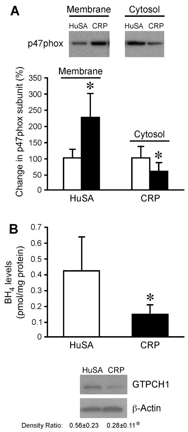
A) Effect of in-vivo CRP treatment on p47phox translocation. Aortic segments were isolated from HuSA-treated and CRP-treated rats and relative protein levels of p47 phox subunit in membrane and cytosol fractions of tissue homogenates were measured. The top panel shows a representative immunoblot using p47phox antibody and the bottom panel shows the densitometric analysis. *P<0.05 vs. HuSA. Data represent 4 rats per group. B) Effect of in-vivo CRP treatment on BH4 levels and GTPCH1 expression. The top panel shows BH4 levels in aortic homogenates isolated from HuSA-treated and CRP-treated rats. The bottom panel shows immunoblots for GTPCH1 and β–actin from these aortic homogenates. The densitometric ratio is provided. Data represent 4 rats per group for both BH4 and GTPCH1 analysis. *P<0.05 vs. HuSA.