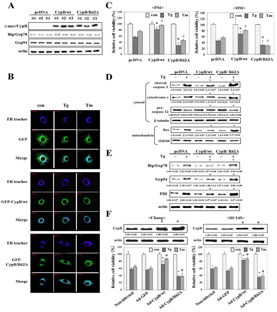
Fig. 3.
Overexpression of cyclophilin B attenuates ER stress-induced cell death. (A) Expression level of CypB/wt or CypB/R62A in the indicated transfectants was analyzed by western blotting. Three different clones in each case were analyzed to exclude the possibility of clonal specificity. CypB was tagged with myc. ER chaperon proteins such as Bip and Grp94 were detected as a reference to monitor the effect of overexpressed CypB on other ER chaperon proteins. Actin was used as a loading control. (B) Localization of overexpressed CypB. After 24 hours of treatment with Tg or Tm under proliferation condition, localization of GFP-CypB/wt or GFP-CypB/R62A in the cells was examined by microscopy. ER tracker was used to identify of the ER. Upper panels shows the localization of GFP alone. Middle panels shows the localization of GFP-CypB/wt, and the lower panels shows the location of GFP-CypB/R62A. ER tracker, blue; GFP, green. Bars, 20 µm. (C) Attenuation of ER stress-induced cell death by overexpressed CypB/wt in proliferation (PM) or differentiation condition (DM) was determined by MTT assay. Note that CypB/R62A overexpression further reduces cell viability. The data are expressed as the means ± s.d. obtained from five independent experiments. *P<0.05 versus Tg-treated pcDNA transfected cells; #P<0.05 versus Tm-treated pcDNA transfected cells in PM and DM, respectively. (D)Western blot with apoptosis marker antibodies: caspase 3 (cleaved form), cytochrome c (cytosolic), procaspase 12 and Bax (mitochondrial). Numbers under each band are representative of at least five different experiments and are expressed as the means ± s.d. *P<0.05 versus Tg-treated pcDNA. (E) Changes in induction of ER resident proteins in CypB/wt and CypB/R62A transfectants. Total cellular extracts from the cells were analyzed for Bip, Grp94 and PDI by western blot. Numbers under each band are representative of at least five different experiments and are expressed as the means ± s.d. *P<0.05 versus Tg-treated pcDNA. (F) The defensive role of CypB is not limited to cell type. Chang and DU145 cells were infected with no adenovirus, Ad-GFP, Ad-CypB/wt, or Ad-CypB/R62A. The expression levels of CypB were detected by western blotting. Their transduction frequency (~60%) was monitored by GFP positivity. Numbers under each band are representative of at least five different experiments and are expressed as the means ± s.d. *P<0.05 versus non-infected Chang and DU145 cells, respectively. The survival rates of each infectant after ER stress were quantified by MTT assay. Ad-GFP, green fluorescence protein adenovirus; Ad-CypB/wt, CypB adenovirus; Ad-CypB/R62A, CypB/R62A adenovirus. The data represent the means ± s.d. obtained from five independent experiments. *P<0.05 versus Tg-treated non-infected cells; #P<0.05 versus Tm-treated non-infected Chang and DU145 cells.

