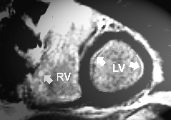
Figure 3.
Magnetic resonance imaging of patient number 6 (short axis) showing a signal enhancement of the septal (arrow) and slightly of the basolateral region (arrow) of the left ventricular myocardium (late enhancement, segmental inversion recovery - TurboFLASH 2D image). RV: right ventricle. LV: left ventricle.

