Figure 14.

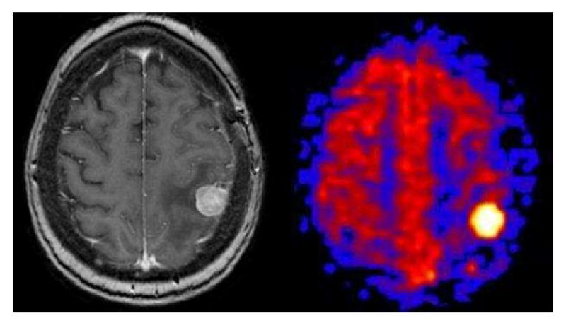
Solid Breast Metastasis. Axial post-contrast T1 weighted image shows a solid enhancing focus in the posterior left frontal lobe with surrounding vasogenic edema. PASL CBF map shows an intensely hyperperfused focus.

Solid Breast Metastasis. Axial post-contrast T1 weighted image shows a solid enhancing focus in the posterior left frontal lobe with surrounding vasogenic edema. PASL CBF map shows an intensely hyperperfused focus.