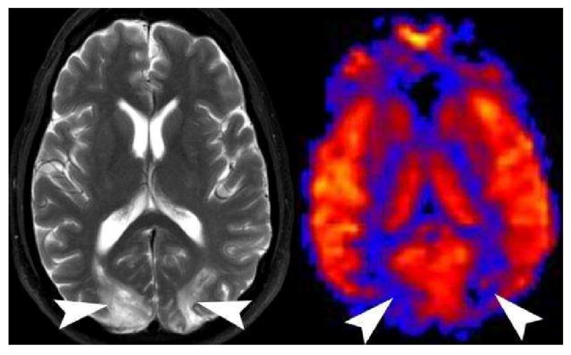
Figure 21.

Posterior Reversible Encephalopathy Syndrome (PRES) on PASL. Axial T2 weighted image shows increased symmetric bilateral signal in the occipital hemispheres (arrowheads). PASL CBF map reveals significant hypoperfusion in the corresponding vascular territories (arrowheads). PRES appears to have a time dependent appearance. Patients imaged acutely tend to have hyperperfusion in the affected territories, while those patients imaged in the subacute phase are hypoperfused.
