
Figure 22.
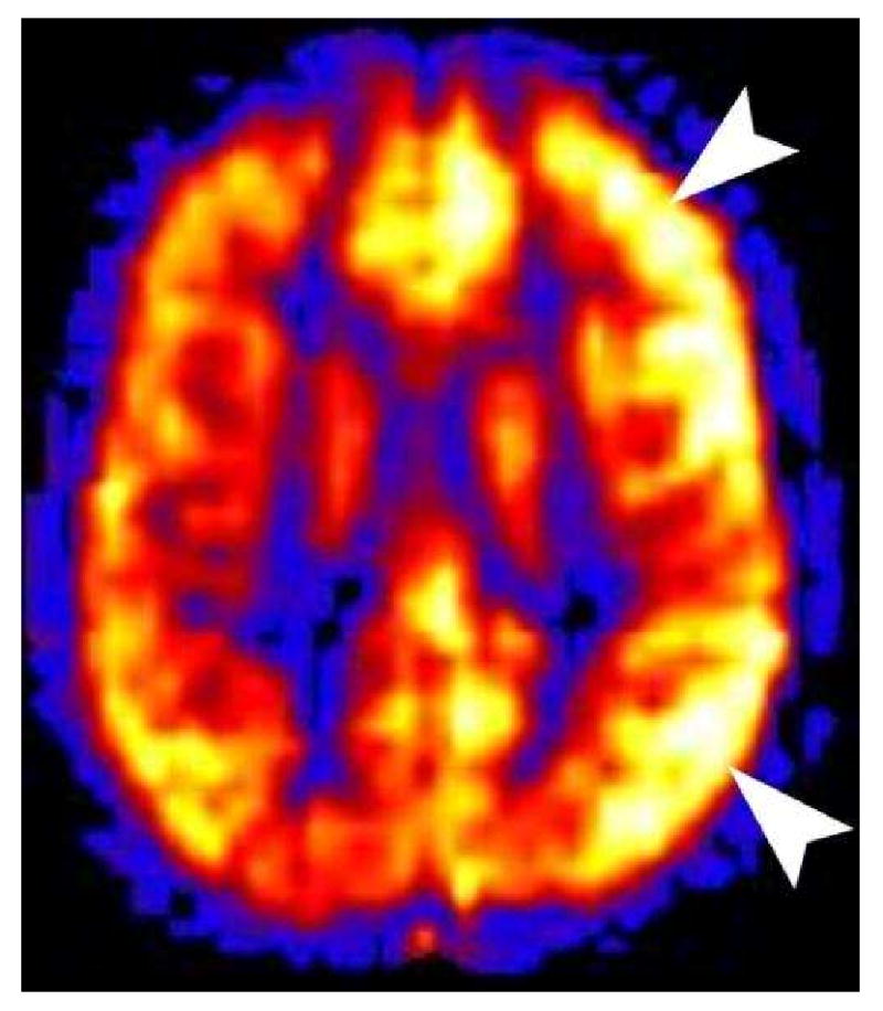
Hemiplegic Migraine. This 11 year old male presented with right hemiplegia followed by a severe migraine headache. All conventional sequences were normal. PASL showed marked hyperperfusion in the left cerebral cortex. Hyperperfusion of the symptomatic cortex is seen in patients imaged during the migraine headache. Hypoperfusion is seen if the patient is imaged during the aura phase.