
Figure 23.
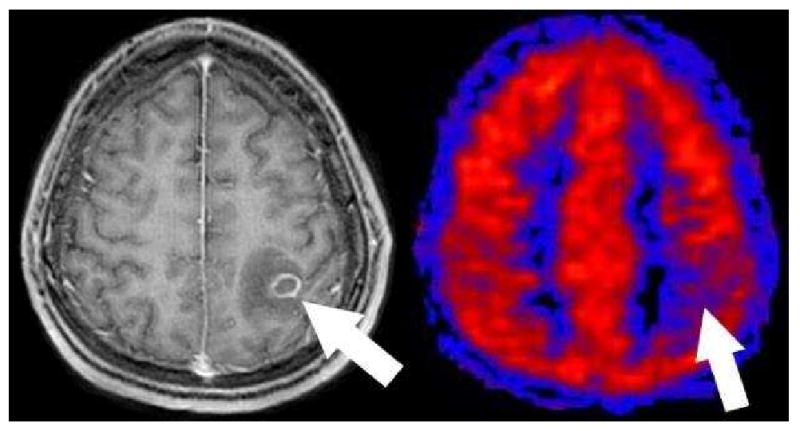
Toxoplasmosis on PASL. Axial post contrast T1 image shows a ring enhancing mass with surrounding edema in this immunocompromised patient (arrow). PASL CBF map shows that the lesion is hypoperfused relative to grey matter. This hypoperfused pattern has been consistently seen in all toxoplasmosis cases to date and may be useful to help distinguish toxoplasmosis from lymphoma.